Peek Into the Hidden World Inside of Cells
Using state-of-the-art equipment, biomedical scientists are answering some of today's biggest questions at the microscopic level.
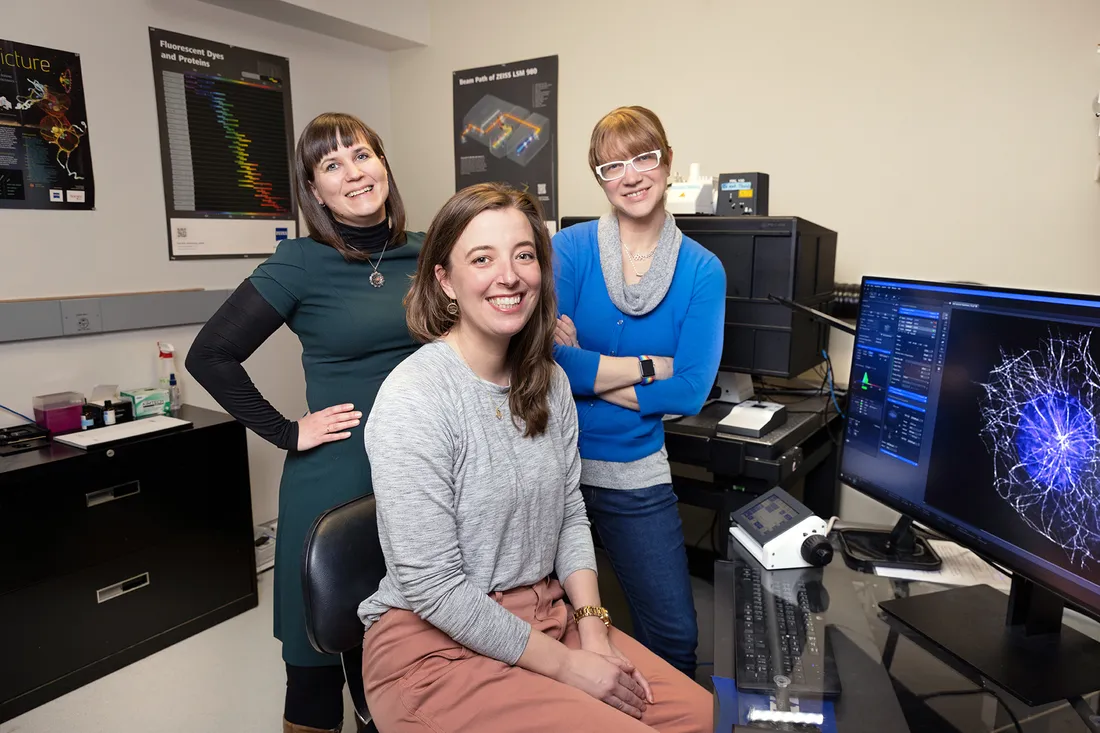
Biology professor Heidi Hehnly flanked by physics professors Mirna Skanata and Jennifer Ross.
It was almost by accident that Antonie van Leeuwenhoek, a 17th-century cloth merchant, first saw living cells. Training his homemade magnifying glass on a drop of pond water, he glimpsed a microbial world teeming with life.
Van Leeuwenhoek called the wormlike creatures “animalcules,” which turned out to be ordinary bacteria. His lenses later tracked the movements of muscle fibers, red blood cells and blood-infused capillaries.
Ironically, the “Father of Microbiology” doubted that technology would ever advance enough to reveal the inner structures of cells—arguably the peak of structural biology.
“We’ve barely scratched the surface of what we know about subcellular activities,” says Heidi Hehnly, a biology professor in Syracuse University’s College of Arts and Sciences. “That’s because cells are highly intricate and, even under controlled conditions, are tricky to work with.”
Microscopy allows us to examine the collective behavior of cells in a variety of biological tissues, from plants to vertebrates. It’s cross-disciplinary research at its best.
Professor Heidi Hehnly
Hehnly oversees the Blatt BioImaging Center on the third floor of the Life Sciences Complex. Using new and emerging technologies, she studies the molecular origins of disease. Her work is emblematic of the center’s mission, which is to serve students, faculty and staff from across campus as well as those at SUNY Upstate Medical University and the SUNY College of Environmental Science and Forestry.
“We offer a range of microscopes and services,” continues Hehnly, alluding to workshops, collaborative research opportunities and instrument rentals. “The more we know about how parts of a cell fit together, the better able they can be engineered for therapeutic purposes.”
Illuminating Possibility
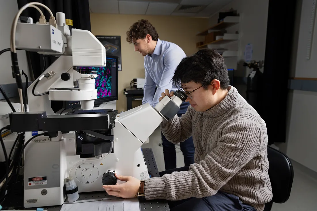
Physics major Hong Beom Lee ’23 operates a Zeiss LSM 710 confocal microscope. Biology major Jonah Da Silva ’24 is in the background.
The Blatt BioImaging Center is a hub for microscopy, which abounds in all corners of campus—from the departments of biology, biomedical and chemical engineering, chemistry, Earth and environmental sciences, and physics to the BioInspired Institute and Forensic and National Security Sciences Institute. Named for retired educator Sydelle Blatt ’51, the three-year-old center houses a fleet of light microscopes (aka scopes) combining visible light and a system of lenses to produce magnified images.
Hehnly uses four different instruments, one of which is a spinning disk confocal scope—named for the hundreds of pinholes arranged in spirals on an opaque disk. When spun, the pinholes scan across a specific optical section of a specimen, building up an image in the process.
“The spinning disk confocal is my workhorse,” she says. “It lets me visualize different complex samples, like zebrafish embryos and human cancer cells, which are model systems for medical research.”
Hehnly is a key member of the BioInspired Institute, where she co-leads the Mechanics of Development and Disease working group with physics professor Alison Patteson. The interinstitutional team develops research strategies for optically sectioning complex samples in real time. In addition to Patteson’s work with swarming bacteria, the group is known for its inquiry into the biomechanics of tissue formation—an area of expertise for Lisa Manning and Jeff Amack, professors of physics and biology, respectively, at Syracuse and SUNY Upstate.
“Microscopy allows us to examine the collective behavior of cells in a variety of biological tissues, from plants to vertebrates,” says Hehnly, who also blends microscopy with art in her Bio-Art Mixer event series. “It’s cross-disciplinary research at its best.”
A Visionary Lens
If anyone can nurture talent, it’s Hehnly, whose students include Jonah Da Silva ’24, a junior majoring in biology. She’s taught him how to image centrioles—barrel-shaped organelles that govern microtubules during cell division. (Microtubules are hollow rods that give cells their shape.) Most cells have at least two centrioles, a mother and a daughter, that reside near the nucleus.
Peering into a Zeiss 980 laser scanning microscope, Da Silva observes a mother centriole interacting with other organelles. Monitoring such activity, he explains, helps him understand how cells grow and migrate as well as contribute to wound healing.
“Dr. Hehnly has shown me how to retrieve the best images possible,” says Da Silva, who plans to enroll in a M.D./Ph.D. program after graduation. “Microscopy adds to my scientific knowledge while fostering a sense of community on campus.”
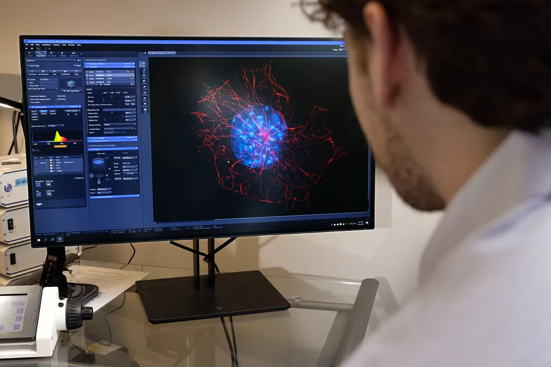
Da Silva observes interactions between microtubules (red) and the nucleus (blue).
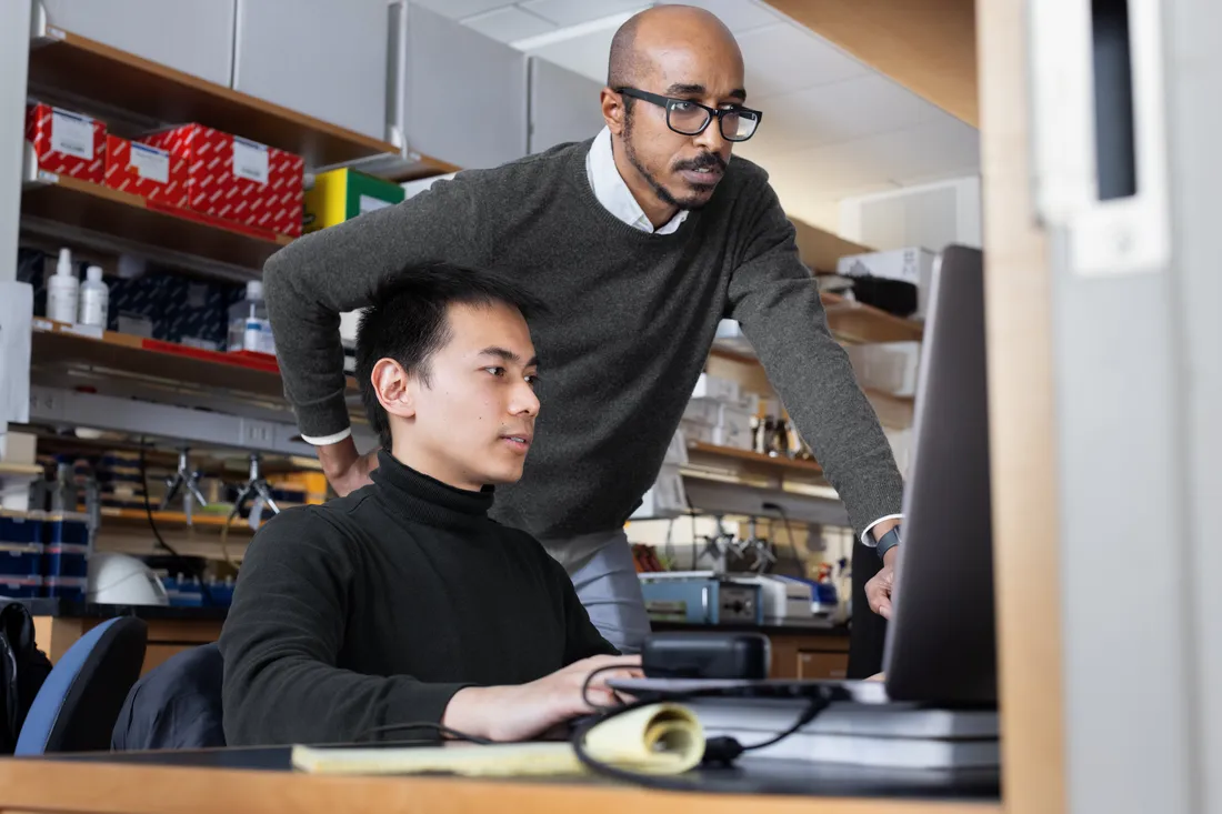
Maxx Swoger, a physics doctoral student, concurs. He studies a structural protein called vimentin to gain fresh insight into cell migration, motility and adhesion.
Sometimes Swoger uses instruments belonging to Hehnly and physics professor Jennifer Ross; other times, he fires up one of Blatt’s super-resolution confocal scopes. His primary method is fluorescent microscopy, where a specimen absorbs a wavelength of light while emitting another.
“I recently figured out how to do a 3D rendering of a nucleus under stress,” Swoger gushes. “I was able to see an indentation, where a bundle of fibers was pushing down on the nucleus. It was amazing.”
The Physics of Discovery
Ross is part of a growing team of researchers working at the nexus of physics and biology. She’s interested in how cells organize their insides without a manager—something involving “a lot of physics,” says the physics chair. “To me, seeing is believing, so we need high-end scopes for what we do.”
Like Hehnly, Ross uses fluorescent microscopy to determine the locations of molecules traveling through space and time. Direct observations are courtesy of Total Internal Reflection Fluorescence (TIRF) microscopy, where lasers bounce off the surface of a sample, creating a short-lived decaying light wave. “The surface is where these molecules are found,” Ross says.
Whereas confocal scopes magnify thick samples, TIRF instruments “see” only thin, dilute ones. Ross tracks and measures each sample’s position until it no longer emits a fluorescent signal. This inability to fluoresce, caused by an overexposure to laser light, is called photobleaching and can occur within seconds.
Microscopy adds to my scientific knowledge while fostering a sense of community on campus.
Jonah Da Silva ’24
Ross’ homemade TIRF scope is indicative of her DIY ethos. She’s currently building optical tweezers to manipulate particles a millionth of a meter in size “It’s like a tractor beam for tiny objects,” says Ross, drawing comparisons to the tools in Star Wars that starships use to reel in another. “And our students get to use it.”
Hong Beom Lee ’23, a senior majoring in physics, is one of Ross’ mentees. He credits her for showing him how to use TIRF microscopy to investigate the self-organization of microtubules, especially when they crosslink, or chemically join, with other protein molecules.
“If she’s taught me anything, it’s that I can learn from my mistakes,” says Lee, who also is mastering the fine art of aligning different microscopic settings, like magnification and resolution, to produce 3D images. “Challenges almost always give rise to opportunities.”
Ideas That Excite
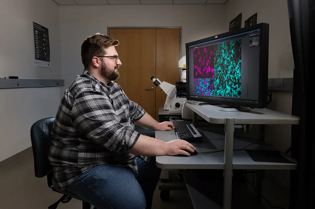
Maxx Swoger, a physics doctoral student, uses the Blatt BioImaging Center to study cell migration, motility and adhesion.
Whereas Ross and Hehnly specialize in molecular and cellular specimens, their physics colleague Mirna Skanata measures signals from “thick, messy, squishy brains” in motion.
Skanata’s research hinges on two-photon imaging, in which quantum-sized energy packets are used to “excite” fluorescent molecules. Such infrared laser-activity lets her optically section the interiors of specimens ranging from tissues and organs to small animals.
Most of her work revolves around the brain of a baby fruit fly. With about 10,000 neurons packed into a space a fraction of the size of a poppy seed, the larval brain is small enough to examine at the cellular level. It’s also big enough to support complex behaviors like learning, smelling and moving.
Skanata develops technologies that enable her to image “circuit-wide” activity in freely moving animals. By studying the link between neural activity and behavior, she seeks to understand how brains compute.
“Even at the larval stage, the fruit fly exhibits a large repertoire of behaviors,” says Skanata, a biophysicist and systems neuroscientist. “Multiphoton microscopy allows me to read and manipulate neural activity in a time-efficient, non-invasive way.”
The implications for understanding processes like learning and memory are “immense,” she adds.
The kind of work that Skanata does, along with Hehnly, Ross and others, more than cultivates a culture of innovation and inquiry. It also contributes to the University’s sustained progress. Leave it to the Blatt BioImaging Center to do what Van Leeuwenhoek once considered impossible—push biomedicine into the realms of the unfathomable.
“Imaging neural activity from freely moving animals is still in its infancy, but it allows us to generate approaches with therapeutic value,” Skanata concludes. “Thanks to the Blatt BioImaging Center, our students are on the front lines, making scientific history.”